
Accordian Closable

Top Skin Analyzer Device Types for Global Buyers?

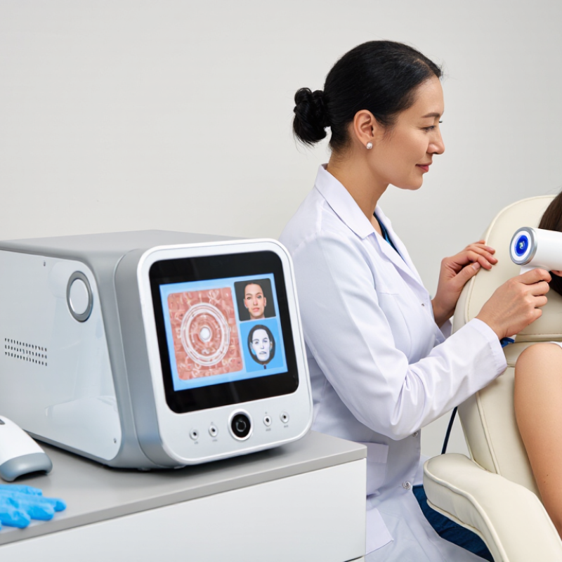
A Skin Analyzer Device can turn a quick consultation into a closer look at pores, pigmentation, texture, and moisture. For global buyers, the challenge is choosing the right device for the setting. A handheld scanner may suit a compact clinic. A tabletop system can offer broader imaging for a busy skincare studio. Some platforms use multiple light modes; others focus on a smaller set of measurements. More features do not automatically mean better decisions.

Dermatologist and cosmetic-science expert Dr. Zoe Diana Draelos is a relevant voice in skin assessment. The following line is an editorial paraphrase, not a verified direct quotation: “Useful skin analysis depends on consistent measurements and careful interpretation.” That distinction matters. A glowing color map can look convincing, yet room lighting, camera distance, and operator technique may change the result. Skin tone and device settings deserve attention, too.

This guide compares common Skin Analyzer Device types by imaging method, workflow, and practical use. It also considers what buyers should verify: repeatability, report clarity, training needs, software support, and the evidence behind each feature. A device should help professionals explain findings, not replace clinical judgment. Some comparisons will remain imperfect, because manufacturers do not always measure the same things in the same way. That is worth remembering before purchase. The best fit is usually the one that produces usable, consistent information in the buyer’s actual workspace—not the one with the longest feature list.
Top Skin Analyzer Device Types for Global Buyers?

How Skin Analyzer Devices Are Classified by Form and Function

Skin analyzers are commonly grouped by their physical form. Handheld units are compact and easy to move between rooms. Some connect to a phone or tablet, while others include a built-in screen. Desktop systems usually have a camera stand, controlled lighting, and a larger display. They suit fixed consultation spaces, but need more counter space. Small devices travel well.

Function offers another useful way to classify them. Imaging-focused systems capture visible-light photographs to show features such as pores, texture, and uneven tone. Some use additional light modes to reveal details that standard images may not show clearly. Probe-based devices measure specific surface conditions, such as moisture or oil levels. They can provide quick readings, though results depend on placement and preparation.

Many devices combine cameras, light sources, and measurement probes. This division is useful, though not perfect. A camera can document appearance, but it does not establish a medical diagnosis. Buyers should check which measurements a device actually provides, how results are displayed, and whether users can repeat readings under similar conditions. Even a clean image can mislead if lighting changes. Keep the room setup consistent, and treat scores as reference points rather than complete assessments.

Portable Skin Analyzers for Mobile Consultations

Portable skin analyzers make consultations easier when clients cannot visit a fixed treatment room. A compact device can travel between consultation spaces, pop-up clinics, or home visits. Small enough to carry. Before scanning, place it on a steady surface and clean the lens. Use consistent lighting and ask clients to remove makeup when appropriate. A rushed image may look precise but still mislead.

Many devices capture close-up images of visible features such as texture, pores, and uneven tone. Treat these results as prompts for discussion, not as a diagnosis. Ask about recent sun exposure, products, and skin sensitivity; each can affect how an image appears. At follow-up visits, keep the camera distance and angle consistent. That matters. Recording the date and scan settings also helps staff compare results without overstating small changes.

For mobile consultations, consider battery life, screen readability, storage, and cleaning needs alongside image features. A bright screen helps near a window, though glare can hide details. Check that images remain clear after transfer and that access and consent procedures suit the clinic’s workflow. Portable tools still have limits. One scan rarely tells the whole story, and an unexpected reading deserves a closer look.

Desktop Systems for Clinics and Beauty Centers

Desktop skin analyzer systems suit clinics and beauty centers that need repeatable consultation images without occupying a treatment room. A stable counter, fixed camera distance, and consistent lighting help staff compare visible features such as pores, pigmentation, and redness over time. The ISAPS 2023 Global Survey recorded about 19.1 million non-surgical aesthetic procedures worldwide. This signals substantial interest in non-surgical services, but it does not measure analyzer sales; treat it as service-demand context, not proof of device-market growth. These systems support cosmetic assessments, not medical diagnosis.

Tips: Check image consistency, report clarity, staff training, and maintenance before purchase. Keep the room lighting steady. Small setup changes can affect comparisons.

For daily use, assess whether the desktop unit fits the consultation flow: Can staff capture images quickly, explain results plainly, and store records securely? Ask suppliers for repeatability information and a live demonstration using varied skin tones. A practical weakness remains: attractive reports may imply more certainty than measurements justify. Lighting, positioning, and user technique still matter. Reflect on whether the device improves conversations, rather than simply adding more numbers.

Top Skin Analyzer Device Types for Global Buyers? - Desktop Systems for Clinics and Beauty Centers
Desktop System Type Imaging Approach Commonly Presented Skin Features Typical Use Buyer Considerations
Standard Visible-Light Facial Imaging Captures facial photographs under consistent, visible illumination, usually from multiple angles. Visible blemishes, uneven tone, pores, fine lines, and general skin appearance. Consultation records, routine progress photography, and explaining cosmetic skincare options. Results depend on consistent camera position, lighting, and image settings. Visible-light photographs do not reveal features below the skin surface.
Cross-Polarized Imaging Uses polarized illumination and a corresponding camera filter to reduce surface glare and emphasize some subsurface color variation. May make redness and areas of pigmentation appear more distinct in images, alongside visible skin features. Comparing facial images and supporting consultations about the appearance of redness or uneven pigmentation. Image appearance depends on the device, lighting, and software. Findings are not a medical diagnosis and should not be treated as a substitute for clinical assessment.
Ultraviolet (UV) Imaging Uses UV illumination and a camera to record skin responses such as fluorescence or contrast under UV light. Some devices display patterns associated with surface oil, sunscreen distribution, or pigmentation contrast. Educational skin consultations and visual demonstrations when the system is designed and labeled for that purpose. Capabilities vary substantially by system. Follow the manufacturer's safety instructions, including any guidance on exposure and eye protection; do not interpret images as a diagnosis.
Multispectral or Multi-Mode Imaging Combines several image modes, which may include visible, polarized, and UV illumination, depending on the system. Can provide multiple views of visible texture, color variation, and selected image patterns in one session. Clinics and beauty centers seeking a broader set of consultation images and standardized before-and-after records. Check exactly which light modes and image outputs are included. Software scores and labels are system-dependent and should be explained as image-based estimates, not diagnoses.
3D Optical Facial Imaging Uses multiple cameras or structured-light techniques to create a three-dimensional surface representation of the face. Facial shape and surface contour; some systems also estimate the appearance of wrinkles or texture. Visualizing facial surface changes and documenting appearance over time in a fixed, repeatable setup. Accuracy depends on capture conditions, positioning, and the system's validated capabilities. A 3D surface model does not show internal tissue or establish a medical condition.
Integrated Desktop Consultation System Combines a fixed camera station, controlled lighting, image capture, and software for viewing or comparing photographs. Features available depend on the installed camera modes and software; often includes facial images and session-to-session comparisons. High-volume consultation workflows where consistent capture, image storage, and client-facing review are priorities. Evaluate workstation footprint, cleaning requirements, image storage and access controls, staff training, and compatibility with local privacy requirements.

Imaging-Based Analyzers for Detailed Skin Assessment

Imaging-based skin analyzers capture close-up views of pores, pigmentation, fine lines, and surface texture. Consistent lighting matters. Cross-polarized and ultraviolet images can reveal features that ordinary room lighting may hide, while repeat scans help track visible changes over time.

For a fair comparison, keep the camera distance, head position, and facial expression consistent. Small differences can distort the result.

The International Agency for Research on Cancer estimated about 330,000 new melanoma cases worldwide in 2022, according to its Global Cancer Observatory. This figure describes disease burden, not analyzer accuracy; imaging scores should not be treated as a diagnosis.

The International Skin Imaging Collaboration’s archive contains more than 70,000 dermoscopic images, illustrating the scale of image data used in skin research. But research images and consumer analyzer readings are not interchangeable.

A bright pigmentation map can look convincing. Still, the numbers may appear more certain than the skin itself. Human review and clear explanations of each device’s limits remain important.

Key Factors Global Buyers Use to Compare Device Types

Global buyers compare skin analyzer device types by the work they need to do, not by feature count alone. Handheld units suit mobile consultations and smaller rooms. Desktop systems often offer a larger screen and guided imaging, but need counter space and staff training. Multi-angle imaging may show texture, pores, and visible pigmentation under consistent lighting. A single measurement can be easier to explain, though it may provide less context.

Check how the device handles different skin tones and lighting conditions. Ask for repeatability data, calibration guidance, and clear limits on what its readings mean.

Skin analysis can support cosmetic consultations; it should not replace a clinical diagnosis. Practical details matter, too: setup time, language options, data storage controls, service access, and replacement parts can affect daily use.

A low purchase price may hide ongoing costs. That deserves caution. Before choosing, test the device with typical users and observe whether staff can capture similar images twice. Results may look precise, yet small changes in distance or room light can alter them.

Buyers should also verify local import, electrical, and data-handling requirements before placing an order.